


Search | Sitemap | Navigation |  |
|
||||||||||||||||||||||||||
|
||||||||||||||||||||||||||
|
||||||||||||||||||||||||||
|


Markus Dahlem

Markus Dahlem
I develop neural network models of visual cortex to model the neurological symptoms of migraine in terms of functional cortical organization. Combined with nonlinear wave dynamics this approach can help to explain what happens to the brain during a migraine attack. Computer models of the process are helping to understand this complex phenomenon.
The main goals of my research are devoted to: (1) understanding cortical susceptibility to episodic focal symptoms in terms of neural circuitry, and (2) using the knowledge of the dynamical state of the circuit to develop novel strategies to prevent such symptoms which not only are very disturbing but also are believed to trigger trigeminovascular activation, pain that is.
Scotoma propagation
Animated migraine scotoma. © 2005 Markus Dahlem
I showed that travelling but spatially confined visual field defects can be explained by nonlinear reaction-diffusion systems in a particular regime of excitability (Dahlem & Müller, 2003, Dahlem & Müller, 2004). The typical localization-related migraine symptoms have a well-delineated focal (or multi focal) neuronal pathology that does not engulf all of posterior cortex as formerly thought.

Schematic views of cortical activity in the human brain in the human brain. Top row is time series of CSD adapted from Lauritzen (1987) with permission. Middle row is the same phenomenon assuming a much lower susceptibility for CSD (Dahlem and Müller, 2003). Bottom row: corresponding typical episodic focal symptoms.
We cannot conclude whether it is spreading depression (SD) or the human equivalent neural reaction-diffusion process. It is known that transient ischaemic attacks (TIA) can be excluded because they have a distinctive sudden onset. However, focal symptoms caused by other vascularly mediated pathophysiologic processes (e.g. ischaemia-induced migraine aura) do not show such a crucial clinical feature, but nonetheless different morphology.
|
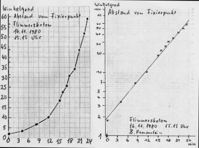
Have you made records of the progression of visual migraine aura? The graph below shows the eccentricity of migraine phosphenes as an example of such data records.
Example of measurements of migraine aura by Bernhard Hassenstein (1980). Please contact Markus Dahlem |
Model of zigzag pattern
To examine the positive symptoms of the typical fortification pattern, i.e., a scintillating zigzag pattern at the leading edge of the scotoma, I basically implemented the suggestion from Richards (1971). Namely that the fortification pattern may be explained with reference to the layout of functional domains in V1, i.e., the orientation map (Dahlem et al., 2000). Based on the responses of ensembles of neurons, a neural population vector (population code) was calculated. Each population was represented in the visual field by an edge with the orientation of the population vector. Population responses may overlap in the visual field, in which case the more dominant population displaces the weaker. This procedure results in a zigzag pattern very similar to those reported by migraine patients.
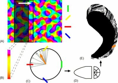
Model of fortification pattern by Markus Dahlem © 2004 Progress in Neurobiology. (cited from Dahlem and Chronicle 2004)
The static appearance of the zigzag is still inconclusive, as similar forms would result from orientation layouts that are arranged in linear parallel stripes. I next animated the zigzag pattern in the expectation that this would reveal further temporal details of the scintillating fortification. The animation was created from the sequence of visual impressions caused by the excitation pattern at subsequent cortical positions. It may be viewed here, but please note that this page include materials that flicker and so may not be suitable for users with migraine with aura or photosensitive epilepsy.
References
Dahlem MA, Chronicle EP. A computational perspective on migraine aura. Prog Neurobiol 2004; 74: 351-361.
Dahlem MA, Müller SC. Reaction-diffusion waves in neural tissue and the window of cortical excitability. Annalen der Physik 2004; 13: 442-449.
Dahlem MA, Müller SC. Migraine aura dynamics after reverse retinotopic mapping of weak excitation waves in the primary visual cortex. Biol Cybern 2003; 88: 419-424.
Dahlem MA, Engelmann R, Löwel S, Müller SC. Does the migraine aura reflect cortical organization? Eur J Neurosci 2000; 12: 767-770.
Richards W. The fortification illusions of migraines. Sci Am 1971; 224: 88-96.
Wiener N, Rosenbluth A. The mathematical formulation of the problem of conduction of impulses in a network of connected excitable elements, specifically in cardiac muscle. Arch Inst Cardiol Mex 1946; 16: 205-265.
Author: Markus Dahlem
Last modification of this page: August 25, 2006
 Top of the page
| · | News |
| · | Medical Professionals |
| · | Medical Studies |
| · | Simulations |
| · | Morphogenesis |
Copyright © 2005 Migraine Aura Foundation, All rights reserved. Last modification of this site: August 25, 2006
Thanks to: RAFFELT MEDIENDESIGN and GNU software | webmaster@migraine-aura.org
http://migraine-aura.org/EN/Markus_Dahlem.html