|
|
|
Imaging Migraine Aura
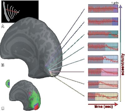
Migraine study with functional magnetic resonance imaging © 2001 PNAS (cited from Hadjikhani et al. 2001)
First experimental evidence for a neural origin in migraine came from the so-called "spreading oligemia" observed in migraine patients using single-photon emission computed tomography (SPECT, see Olesen et al., 1990). It was not an obvious step, however, to conclude that this experimental observation can be equated with "cortical spreading depression" (CSD), the neural disorder in question. Quite the contrary, Olsen (1995) argues that the spreading oligemia observed with SPECT techniques is merely an artifact and that the ordered sequence of aura symptoms can also be explained by a gradient in susceptibility to ischaemia (a vascular disorder). In that case, although the aura symptoms appear as being caused by a traveling cortical phenomenon, in fact a steady cortical gradient merely coordinates wave-like behavior of a transient ischemic attack (TIA). The principal objections to the vascular theory of the aura arose from the well matched spatio-tmporal pattern of the symptoms to CSD characteristics. Furthermore, the spreading disorder does not seem to respect vascular territories.
Today, the conflicting experimental evidence is challenged by further non-invasive imaging methods, such as functional Magnetic Resonance Imaging (fMRI). These investigations may let us conclude, that both neural and vascular mechanism play a pathogenetic role in migraine aura. Many researches have contributed to this field in the last years. More details about their work will appear here soon.
References
Friberg L, Olesen J, Lassen N A, Olsen T S, Karle A. Cerebral oxygen extraction, oxygen consumption, and regional cerebral blood flow during the aura phase of migraine. Stroke 1994; 25: 974-979.
Hadjikhani N, Sanchez Del Rio M, Wu O, Schwartz D, Bakker D, Fischl B, Kwong KK, Cutrer FM, Rosen BR, Tootell RB, Sorensen AG, Moskowitz MA. Mechanisms of migraine aura revealed by functional MRI in human visual cortex. Proc Natl Acad Sci USA 2001; 98: 4687-4692.
Lauritzen M. Pathophysiology of the migraine aura. The spreading depression theory. Brain 1994; 117: 199-210.
Olesen J, Friberg L, Olsen TS, Iversen HK, Lassen NA, Andersen AR, Karle A. Timing and topography of cerebral blood flow, aura, and headache during migraine attacks. Ann Neurol 1990; 28: 791-798.
Olsen TS. Pathophysiology of the migraine aura: the spreading depression theory. Brain 1995; 118: 307-308.
Author: Markus Dahlem
Last modification of this page: Tuesday January 31. 2006
 Top of the page Top of the page


|
|
|