


Search | Sitemap | Navigation |  |
|
||||||||||||||||||||||||||
|
||||||||||||||||||||||||||
|
||||||||||||||||||||||||||
|


| James A. Reggia |
Otto-Joachim Grüsser
Otto-Joachim Grüsser (1932-1995) at the Berlin State Library in the early 90th.
A remarkable career in neurology, ophthalmology, neuroscience, perception, and the history of these disciplines made Otto-Joachim Grüsser an renowned researcher. He started with medical studies and after he completed the preclinical education at the University of Tübingen, he continued 1954 in Freiburg. His superviser, Richard Jung, also worked on migraine aura. But that was many years after Grüsser had visited his lab. Jung's first migraine attack with visual aura was not before 1960 and he published 19 years later a remarkable paper on effects of vestibular stimulation on visual aura (Jung, 1979, see also here).
Grüsser first published a Society of Neuroscience abstract about the aura in 1991 and later a more detailed analysis (Grüsser, 1995), in particular about the functional mapping of the human retina (known as retinotopy) onto the primary visual cortex (V1).
His main result is that the speed of his visual symptoms is exponentially increasing towards the periphery. This is in good agreement with the assumtion that the cortical speed is constant (see also here) and that retinal polar coordinates are conformally mapped to cartesian cortical coordinates. The latter assumption is often refered to as complex logarithmic mapping, but maybe better interpreted as the requirement of isotropic retino-cortical magnification.
There is one further hidden assumption though. He observed a large interindividual variability in the magification factors, which he attributes to the large variability of overall V1 area as reported by Stensaas et al. (1974) and more recently confirmed by Andrews et al. (1997). Today, with modern imaging such as functional magnetic resonance imaging (fMRI), one could provide a detailed meassure of V1 area and assert this assumption.
Grüsser also made detailed suggestion why there are zig-zag patterns in the fortification. As Richards (1971) he thinks that cortical orientation hypercolumns are the basic elements of the zig-zag pattern. To explain the peculiar shape he argues along the lines of Schwartz (1980) but proposes some new mechanistic steps: "the general rise in neural excitation due to increased potassium concentration along the stripe of Gennari (see below) leads to an increased activation of layer IV neurons, which is modified by lateral inhibitory and excitatory mechanism acting mainly in cell tiers above and below layer IV".

The primary visual cortex (V1) consists of six layers. It contains a prominent stripe of white matter in its layer 4 − the "stripe of Gennari" consisting of the myelinated axons. For this reason, V1 is also referred to as the striate cortex. © Markus Dahlem 2005
Raised potassium concentration and its higher apparent diffusion coefficient along the stripe of Gennari is central in Grüsser's argument. He suggests that "most likely akin to the pottasium diffusion during cortical spreading depression [...] diffusion takes place chiefly within the stripe of Gennari". However, blaming cytoarchitectonic structures introduces inconsistencies. He asks "why migraine symptoms in some patients affect cortical regions far outside area V1". He thinks that "some pathophysiological mechanisms such as spread of neurotransmitters acting on vascular smooth muscle transgress the area V1 border into neighboring cortical areas. This would [also] explain [...] why migraine symptoms [...] eventually evoke severe headache".
The confined appearance of the symptoms in cortical, respectively sensory space is indeed a central question. With functional magnetic resonance imaging this question was not yet sufficiently resolved because all posterior cortex − including several retinotopically organized visual areas − showed simultaneous activation during much of the period of the aura (Hadjikhani et al. 2001). Wilkinson (2004) suggested two alternative explanations. The activation due to spreading depression may despite the fMRI data be limited in extent and "the rest of the spreading activation in adjacent cortical areas represents synaptic activation through feed-forward and feedback circuitry", or alternatively, "the spreading depression process engulfs all of posterior cortex, but that only a subset of this activation results in visual awareness".
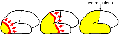
Lateral views of human brain with different time intervals after start of attack. Adapted from Lauritzen (1987) with permission.
The latter alternative is also suggested by a drawing from Lauritzen (1987, 1995), showing a wave moving from the back of the cerebral cortex (occipital pole) toward the front untill it reaches the central sulcus. Lauritzen spearheaded the spreading depression theory of migraine aura. However, it is debatable whether this image is supporting the CSD theory. It is hard to believe that a CSD wave engulfs all of posterior cortex during a migraine attack. Why should only a subset of the activation results in visual awareness and why are migraine symptoms not much more severe when CSD engulfs all of posterior cortex?
It was Grüsser's foresight that recognized the need of a plausible answer to this central question of the spreading depression theory. Dahlem and Müller (2003, 2004, see also the next but one section) recently suggested that physiological parameters are in a specific window of cortical excitabiliy in which the activation pattern are confined in space and time. In this window of excitability, there is an unstable wave segment with a particular size and shape, which will contract tangentially at its free ends and eventually disappear (for a theoretical analysis, see Zykov and Showalter, 2005). Such particle-like waves can explain the limited appearance of aura symptoms without cytoarchitectonic heterogeneities but still consistent with the spreading depression theory.
References
Andrews TJ, Halpern SD, Purves D. Correlated size variations in human visual cortex, lateral geniculate nucleus, and optic tract. J Neurosci 1997; 17: 2859-2868.
Dahlem MA, Müller SC. Migraine aura dynamics after reverse retinotopic mapping of weak excitation waves in the primary visual cortex. Biol Cybern 2003; 88: 419-424.
Dahlem MA, Müller SC. Reaction-diffusion waves in neural tissue and the window of cortical excitability. Annalen der Physik 2004; 13: 442-449.
Grusser OJ. Migraine phosphenes and the retino-cortical magnification factor. Vision Res 1995; 35: 1125-1134.
Grusser OJ, Grusser-Cornehls U. Quantitative studies in migraine phosphenes. Society for Neuroscience Abstracts 1991; 17: 1568.
Grusser OJ. Further studies in migraine phosphenes. Perception 1992; 21 (Suppl 2): 37-38.
Hadjikhani N, Sanchez Del Rio M, Wu O, Schwartz D, Bakker D, Fischl B, Kwong KK, Cutrer FM, Rosen BR, Tootell RB, Sorensen AG, Moskowitz MA. Mechanisms of migraine aura revealed by functional MRI in human visual cortex. Proc Natl Acad Sci USA 2001; 98: 4687-4692.
Jung R. Translokation corticaler Migrainephosphene bei Augenbewegung und vestibularen Reizen. Neuropsychologia 1979; 17: 173-85.
Lauritzen M. Pathophysiology of the migraine aura. The spreading depression theory. Brain 1995; 117: 199-210.
Lauritzen M. Cerebral blood flow in migraine and cortical spreading depression. Acta Neurol Scand Suppl. 1987; 113: 1-40. Review.
Richards W. The fortification illusions of migraines. Sci Am 1971; 224: 88-96.
Schwartz EL. A quantitative model of the functional architecture of human striate cortex with application to visual illusion and cortical texture analysis. Biol Cybern 1980; 37: 63-76.
Stensaas SS, Eddington DK, Dobelle WH. The topography and variability of the primary visual cortex in man. J Neurosurg 1974; 40: 747-55.
Wilkinson F. Auras and other hallucinations: windows on the visual brain. Prog Brain Res 2004; 144: 305-320.
Zykov VS, Showalter K. Wave front interaction model of stabilized propagating wave segments. Phys Rev Lett 2005; 18:068302.
Author: Markus Dahlem
Last modification of this page: Montag Juni 20. 2005
| James A. Reggia |
 Top of the page
| · | News |
| · | Medical Professionals |
| · | Medical Studies |
| · | Morphogenesis |
Copyright © 2005 Migraine Aura Foundation, All rights reserved. Last modification of this site: August 25, 2006
Thanks to: RAFFELT MEDIENDESIGN and GNU software | webmaster@migraine-aura.org
http://migraine-aura.org/EN/Otto-Joachim_Gruesser.html