


Search | Sitemap | Navigation |  |
|
||||||||||||||||||||||||||
|
||||||||||||||||||||||||||
|
||||||||||||||||||||||||||
|


| Neural aura pattern |
What happens Where?
The morphology of the aura symptoms and the morphology of the underlying pathophysiological processes are not the same. Loosely speaking, the spatio-temporal activity pattern of the pathophysiological process is translated by one of the many feature maps in the cerebral cortex into a hallucinatory perception pattern.
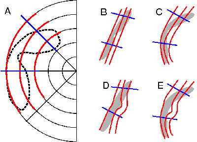
Cortical feature mapsThe cerebral cortex has the topology of a 2-D sheet and is divided into a large number of different areas with topographic maps, also called feature maps. Cortical feature maps are representations of the environment. The topography refers to the fact, that similar features activate nearby locations in the map. Examples are retinotopic, tonotopic, and somatotopic maps. In 2000 Dahlem et. al asked whether the migraine aura reflects such cortical feature maps and provided answers to this question (Dahlem and Müller 2003, Dahlem and Chronicle 2004) based on neural network models. |
Which feature map is involved depends on the cortical area the disturbance takes place. In other words, the morphology of the symptoms does not depend solely on the pathophysiology but also on the feature map. Therefore the morphology of the symptoms does not lead to a taxonomy that is exclusively based on etiology. For a usefull therapeutic taxonomy, we need to consider two factors. First, what happened in the cortex and, second, in which cortical area took it place. Both the "Where" and "What" influence aura morphology and the effects are hard to tell apart.
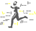
Visualization of the mapping problem: What happens Where? © 2005 Markus Dahlem
While the "What" question refers to the aura etiology, the "Where" question addresses the different feature maps of the cortical areas. Of course, one can try to circumvent both questions. Instead of looking at the aura symptoms, one can record with non-invasive imaging directly the cortical activity during a migraine attack. However, this is only feasible if a patient can trigger the aura. Furthermore, answering these two questions will lead to a better understanding of human cortical feature maps. It is worth to overtaken the obstacles and try to create a taxonomy based on aura symptoms.
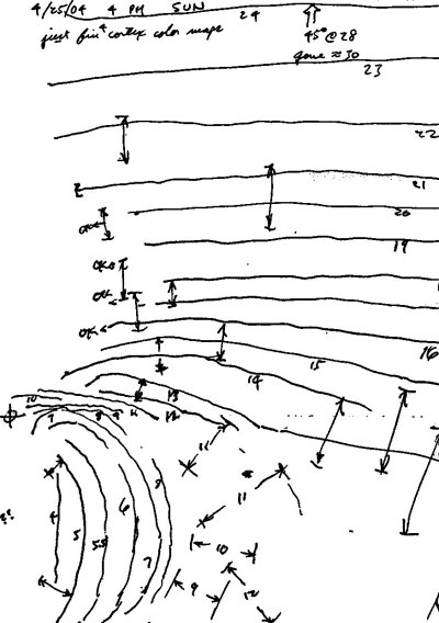
A precise drawing of the spatio-temporal development of the visual impairment. By courtesy of Paul VanValkenburgh. © 2005 Paul VanValkenburgh
A road map to a taxonomy based on visual aura symptoms would be as follows (see also Classification of visual disturbances). We need precise drawings of the spatio-temporal development of the visual impairment during the aura, preferably with computer-assisted diagnostic procedures, as described in the next section. Then, we need to know precisely how so-called retinotopic maps are laid out in the various visual areas of the cortex. Last but not least, we need to know which cortical excitation pattern correspond to a neural and which to a vascular disorder.
References
Dahlem MA, Chronicle EP. A computational perspective on migraine aura. Prog Neurobiol 2004; 74: 351-361.
Dahlem MA, Müller SC. Migraine aura dynamics after reverse retinotopic mapping of weak excitation waves in the primary visual cortex. Biol Cybern 2003; 88: 419-424.
Dahlem MA, Engelmann R, Löwel S, Müller SC. Does the migraine aura reflect cortical organization? Eur J Neurosci 2000; 12: 767-770.
Author: Markus Dahlem
Last modification of this page: Thursday August 25. 2005
| Neural aura pattern |
 Top of the page
| · | News |
| · | Medical Professionals |
| · | Medical Studies |
| · | Classification of visual disturbances |
| · | Neurophysiological mechanisms |
| · | Network models |
Copyright © 2005 Migraine Aura Foundation, All rights reserved. Last modification of this site: August 25, 2006
Thanks to: RAFFELT MEDIENDESIGN and GNU software | webmaster@migraine-aura.org
http://migraine-aura.org/EN/What_happens_Where.html